Facial Analysis and Cephalometrics
Understanding the guiding principles involved in facial analysis and cephalometrics is essential during facial plastic and reconstructive surgery encounters. It is important to remember that each subunit and facial structure is assessed in relation to each other in order to evaluate position, proportion, and ideal aesthetics. This is also important to consider postoperatively as any alteration to a facial subunit or angle could lead to subtle or dramatic changes to one’s physical appearance. Facial proportions and angles vary by age, gender, and ethnicity. Societal and cultural trends may influence the cosmetic goals of a patient. Preoperative and postoperative photographic documentation can aid in patient discussions and assist the surgeon with surgical planning.
- Perform a detailed facial analysis
- Identify the soft tissue and bone anatomic landmarks of the face
- Review the facial subunits
- Recognize the ethnic and gender variations involved in facial analysis
- Explain the standardization of preoperative and postoperative photographic documentation
Anatomy
- Recognize the anatomic soft tissue and bony facial landmarks involved in facial analysis
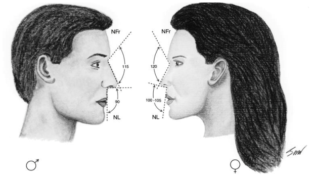
- Identify the cephalometric angles used within facial analysis assessments
- Define the nasofrontal angle, nasofacial angle, nasolabial angle, nasomental angle, auriculocephalic angle, mentocervical angle, and cervicomental angle
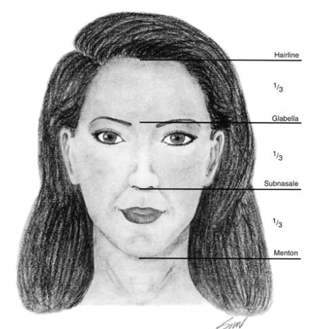
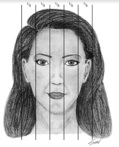
- Describe facial symmetry as it relates to the horizontal thirds and vertical fifths
- Understand the anatomy involving the facial subunits
- The forehead, eyes, nose, cheeks, lips, and chin compose the main facial subunits
- Define the components of a nasal analysis
- Identify the nasal subunits
- Describe the methods used for assessing nasal projection
- Define Goode method
- Define Crumley-Lanser method
- Define Simons’ method
- Define the ratio of the nasal ala-tip
- Define columellar show
- Describe the ideal position of the chin
- Identify the zero meridian of Gonzalez-Ulloa
- Define the ideal lip proportions and aesthetic subunits of the lip
- Define the intercanthal distance
- Describe the location of the supratarsal crease of the upper eyelid and ethnic variations
- Identify the location of the ideal brow positioning and shape in a male and female patient
- Define the components of a nasal analysis
- Understand the relationship of the neck as well as the ears in complete facial analysis
- The forehead, eyes, nose, cheeks, lips, and chin compose the main facial subunits
- Recognize the variations in facial aesthetics related to ethnicity
- Zimbler M. Aesthetic Facial Analysis. In: Cummings Otolaryngology Head and Neck Surgery. 6th Ed. Philadelphia, PA: Saunders; 2015. p237-284
- Papel, I. Facial Analysis and Nasal Aesthetics. Aesth. Plast. Surg. 26, S13 (2002). https://doi-org.ezproxy.bu.edu/10.1007/s00266-002-4317-3
- Bueller H. Ideal Facial Relationships and Goals. Facial Plast Surg. 2018; 34(5):458‐465. doi:10.1055/s-0038-1669401
- Cohen MB, Ezzat WH. Aesthetic Facial Analysis. In: Sataloff's Comprehensive Textbook of Otolaryngology: Head and Neck Surgery: Facial Plastic and Reconstructive Surgery. 1st Ed. Philadelphia, PA: Jaypee Brothers Medical Publishers; 2016. p. 101-113
- Bergman RT. Cephalometric soft tissue facial analysis. Am J Orthod Dentofacial Orthop. 1999;116(4):373‐389. doi:10.1016/s0889-5406(99)70222-2
Pathogenesis
- Identify the basis of attractiveness
- Facial proportions and symmetry
- Cultural influences
- Aging process
- Bueller H. Ideal Facial Relationships and Goals. Facial Plast Surg. 2018;34(5):458‐465. doi:10.1055/s-0038-1669401
- Forte AJ, Andrew TW, Colasante C, Persing JA. Perception of Age, Attractiveness, and Tiredness After Isolated and Combined Facial Subunit Aging. Aesthetic Plast Surg. 2015;39(6):856-869. doi:10.1007/s00266-015-0553-1
Patient Evaluation
- Begin with a generalized analysis of patient by assessing age, gender, ethnicity, and hair
- Understand the importance of skin analysis
- Define Fitzpatrick skin type classification
- Define Glogau photoaging classification
- Recognize the facial angles and its overall contribution to the facial analysis
- Describe the facial measurements that contribute to facial symmetry
- Describe nasal projection and rotation
- Understand the importance of communicating with the patient to learn their cosmetic goals
- Bueller H. Ideal Facial Relationships and Goals. Facial Plast Surg. 2018;34(5):458‐465. doi:10.1055/s-0038-1669401
- Zimbler M. Aesthetic Facial Analysis. In: Cummings Otolaryngology Head and Neck Surgery. 6th Ed. Philadelphia, PA: Saunders; 2015. p237-284
- Cohen MB, Ezzat WH. Aesthetic Facial Analysis. In: Sataloff's Comprehensive Textbook of Otolaryngology: Head and Neck Surgery: Facial Plastic and Reconstructive Surgery. 1st Ed. Philadelphia, PA: Jaypee Brothers Medical Publishers; 2016. p. 101-113
Imaging
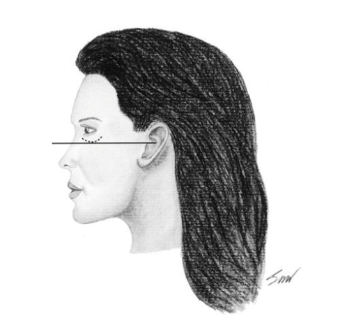
- Define the Frankfort horizontal plane
- Recognize the importance of photographic documentation during pre-operative and postoperative assessments
- Describe patient positioning for standardized photographs
- Identify the advantages and disadvantages to computer generated images
- Zimbler M. Aesthetic Facial Analysis. In: Cummings Otolaryngology Head and Neck Surgery. 6th Ed. Philadelphia, PA: Saunders; 2015. p237-284
- Nair AG, Santhanam A. Clinical Photography for Periorbital and Facial Aesthetic Practice. J Cutan Aesthet Surg. 2016;9(2):115‐121. doi:10.4103/0974-2077.184047
Pathology
- Describe the aging effects noted during facial analysis
- Identify the age related changes of the upper face, midface, and lower face
- Cohen MB, Ezzat WH. Aesthetic Facial Analysis. In: Sataloff's Comprehensive Textbook of Otolaryngology: Head and Neck Surgery: Facial Plastic and Reconstructive Surgery. 1st Ed. Philadelphia, PA: Jaypee Brothers Medical Publishers; 2016. p. 101-113
- Bueller H. Ideal Facial Relationships and Goals. Facial Plast Surg. 2018;34(5):458‐465. doi:10.1055/s-0038-1669401
Case Studies
Case 1:
A 53 year old female presents for a cosmetic consultation and preoperative photographs are obtained. Describe standard patient positioning and photographic views.
- Hair should be placed behind the ear and not impeding the view of any facial features
- Patient will be positioned in anatomic position and in the Frankfort horizontal plane
- Obtain frontal, left oblique, right oblique, left lateral and right lateral views. Can also obtain basal view.
References:
- Nair AG, Santhanam A. Clinical Photography for Periorbital and Facial Aesthetic Practice. J Cutan Aesthet Surg. 2016;9(2):115‐121. doi:10.4103/0974-2077.184047
- Cohen MB, Ezzat WH. Aesthetic Facial Analysis. In: Sataloff's Comprehensive Textbook of Otolaryngology: Head and Neck Surgery: Facial Plastic and Reconstructive Surgery. 1st Ed. Philadelphia, PA: Jaypee Brothers Medical Publishers; 2016. p. 101-113 (Fig N1.)
Case 2:
A 64 year old male presenting for cosmetic consultation. After the patient describes his aesthetic concerns, you notate your clinical and physical assessment of the patient.
- Your assessment begins with a general assessment of appearance, facial symmetry, stated age versus perceived age, hair style, hairline location, ethnicity, gender, skin quality, followed by assessment of each individual subunit
References:
- Cohen MB, Ezzat WH. Aesthetic Facial Analysis. In: Sataloff's Comprehensive Textbook of Otolaryngology: Head and Neck Surgery: Facial Plastic and Reconstructive Surgery. 1st Ed. Philadelphia, PA: Jaypee Brothers Medical Publishers; 2016. p. 101-113
Case 3:
A 36 year old male is concerned about the appearance of his chin and feels that it does not “fit his face”
- Perform a complete facial analysis
- Evaluate his chin in relation to the surrounding facial subunits including the lips and the nose
- Identify the pogonion which is the most anterior soft tissue landmark on the chin
- Determine the zero meridian of Gonzalles-Illoa
- Assess the patient for mandibular dimorphism
References:
- Arroyo HH, Olivetti IP, Lima LF, Jurado JR. Clinical evaluation for chin augmentation: literature review and algorithm proposal. Braz J Otorhinolaryngol. 2016;82:596---601
Review
- Define the Frankfort horizontal line.
- Outline the facial soft tissue anatomic landmarks.
- Describe the subdivision of the lower third of the face.
- What is the ideal brow position in a woman?
- What are the relaxed skin tension lines?
- Zimbler M. Aesthetic Facial Analysis. In: Cummings Otolaryngology Head and Neck Surgery. 6th Ed. Philadelphia, PA: Saunders; 2015. p237-284
- Cohen MB, Ezzat WH. Aesthetic Facial Analysis. In: Sataloff's Comprehensive Textbook of Otolaryngology: Head and Neck Surgery: Facial Plastic and Reconstructive Surgery. 1st Ed. Philadelphia, PA: Jaypee Brothers Medical Publishers; 2016. p. 101-113
Learner must Sign In to access AAO-HNSF education activities.
- Otolaryngology Patient Scenarios (OPS):