Secondary Rhinoplasty
Rhinoplasty surgery is a technically difficult and physically demanding surgery.
You must interact with anatomical considerations such as skin, bone and cartilage in a three-dimensional fashion. Youi must also take into consideration social and cultural norms and appearance. The most common cause of secondary rhinoplasty is Primary rhinoplasty. It is important to conduct a thorough analysis preoperatively and to asses underlying asymmetries or potential pitfalls that can lead to a secondary rhinoplasty. During the informed consent process, it is important to discuss the possibility of a secondary rhinoplasty. This outline describes the most common deformities and how to address them.
- Recognize the indications for Secondary Rhinoplasty.
- List the most common deformities associated with need for secondary rhinoplasty.
- Describe the causes of various deformities associated with need for secondary rhinoplasty.
- Review various methods to correct deformities during a secondary rhinoplasty.
- Explain functional and cosmetic deformities associated with need for secondary rhinoplasty surgery.
- Cite ways to avoid complications during Primary rhinoplasty that can lead to Secondary rhinoplasty.
- Describe three anatomical regions of nose and most common deformities seen in each region.
Anatomy
- Review the Anatomic regions of nose:
- Nasal Bones (Bony Vault) - bony vault is a pyramidal structure that forms the principle structural base. The lateral walls consist of the frontal processes of the maxilla and the nasal bones with union to the frontal bone in the midline.
- Middle Vault
- Upper lateral cartilages (ULC) and Septum
- Nasal Tip
- Lower lateral cartilages
- Medial and lateral crura
- Explain Functional Anatomy of Nose:
- Internal Nasal Valve - formed by caudal edge of ULC, septum and nasal floor. Ideally 15 degrees
- External Nasal Valve - region caudal to the internal valve bounded superolaterally by the caudal edge of the upper lateral cartilage, laterally by the nasal alar and bony piriform aperture of the maxilla, and medially by the septum and columella.
- Septum - the bony and cartilaginous septum is formed by the perpendicular plate of the ethmoid, the vomer, the palatine crest and the quadrangular cartilaginous septum.
- Nasal Turbinates
- Tardy, ME. Rhinoplasty: the art and science. Philadelphia; W.B. Saunders; 1997
- Toriumi DM, Johnson CM Jr: Open structure rhinoplasty. Featured technical points and long-term follow-up. Facial Plastic Surg Clin N Am 1(1):1-22, 1993
- Azizzadeh B, Murphy MR, Johnson CM, Numa W. Master Techniques in Rhinoplasty. Philadelphia; Elsevier Saunders; 2011
Pathogenesis
- Explain most common deformities found in each Anatomic Region of Nose:
- Upper Third
- Rocker deformity
- Asymmetry of nasal bones
- Over-resection of Nasal Bones
- Open Roof Deformity
- Stair Step Deformity
- Middle Third
- Polly Beak Deformity
- Saddle Nose Deformity
- Inverted V deformity
- Dorsal Septal Deflection
- Lower Third
- Bossae
- Pinched Tip
- Nostril Asymmetries
- Alar-Columellar Disproportion
- Visible Grafts
- Poorly Defined Tip
- Columellar Scarring
- Caudal Septal Deflection
- Upper Third
- Describe Functional Complications than can lead to a need for secondary rhinoplasty:
- Nasal Obstruction
- External Nasal Valve Collapse
- Internal Nasal Valve Collapse
- Septal Deviation
- Intranasal Synechia
- Turbinate Hypertrophy
- Nasal Obstruction
- Gaith S. Secondary Rhinoplasty. Indian J Plast Surg. 2008 Oct; 41(Suppl): S80–S87
- Surowitz JB, Most S. Complications of Rhinoplasty. Facial Plast Surg Clin North Am. 2013 Nov;21(4):639-51
Incidence
- Overall revision rate for Primary Septorhinoplasty leading to Secondary Rhinoplasty was 3%
- Revision rate for Secondary Rhinoplasty leading to additional Rhinoplasties- 11%
- Patient Characteristics leading to Secondary rhinoplasty
- Younger age
- Female sex
- Autoimmune defects
- Cosmetic Deformities
- Congenital Deformities
- Spataro E, MD, Piccirillo JF, MD, Kallogjeri D, MD, MPH, et al. Revision Rates and Risk Factors of 175 842 Patients Undergoing Septorhinoplasty. JAMA Facial Plast Surg. 2016;18(3):212-219
Patient Evaluation
Summarize the basic principles in Nasal Analysis:
- Nasal Anatomy
- Nasal Proportions
- Frankfort Horizontal Plane
- Nasofrontal Angle
- Nasomental Angle
- Goode’s Ideal Tip Projection
- Vertical Fifths and Horizontal Thirds of face
- Nasal Subunits
- Frankel A, Mehta U. Nasal Analysis. In: Master Techniques in Rhinoplasty. Philadelphia: Elsevier Saunders; 2011. p. 31-42.
Measurement of Functional Status
Explain how to evaluate for nasal valve collapse and nasal obstruction:
- Cottle Maneuver and Modified Cottle Maneuver
- Lateral wall insufficiency
- Rhee JS, Weaver EM, Park SS, Baker SR, Hilger PA, Kriet JD, Murakami C, Senior BA, Rosenfeld RM, DiVittorio D. Clinical consensus statement: diagnosis and management of nasal valve compromise. Otolaryngol Head Neck Surg. 2010;143:48–59.
- Vaezeafshar R, Moubayed SP, Most SP. Repair of Lateral Wall Insufficiency. JAMA Facial Plast Surg. 2018 Mar 1;20(2):111-115.
Imaging
- Explain importance of Photography to Assess preoperative and postoperative changes associated with Rhinoplasty.
- Identify the use of Computer tomography to evaluate bony and cartilaginous deformities that can lead to need for secondary rhinoplasty.
- Frankel A, Mehta U. Nasal Analysis. In: Master Techniques in Rhinoplasty. Philadelphia: Elsevier Saunders; 2011. p. 31-42.
Treatment
- List the various techniques used to restore form and function in secondary rhinoplasty.
- Surowitz JB, Most S. Complications of Rhinoplasty. Facial Plast Surg Clin North Am. 2013 Nov;21(4):639-51
Surgical Therapies
- Describe various surgical options available for most common causes of Secondary Rhinoplasty:
- Upper Third (bony vault)
- Percutaneous osteotomies - Rocker deformity, stair step deformity, asymmetry of bony vault
- “Double lateral” osteotomy to correct stair step
- Lateral osteotomies to close open roof
- Middle Third
- Polly Beak Deformity- cartilaginous dorsal reduction to that of bony dorsum. Consider Kenalog Injection if scar formation
- Inverted V deformity- Revision with use of spreader grafts (if upper lateral cartilage present), possible onlay crushed cartilage camouflage grafts, consider osteotomies
- Saddle Nose- Revision with dorsal onlay camouflage graft (minor cosmetic deformity) and rib cartilage graft reconstruction (severe deformities)
- Lower Third
- Nasal Tip Deformities
- Bossae- Revision with structural grafting of lateral crura (strut grafts), crushed cartilage, and/or temporalis fascia camouflage graft. Consider excision and closure in limited cases with placement of overlying graft.
- Pinched tip- Removal/revision of any offending tip sutures, possible lateral crural strut grafting, possible repositioning of lateral crura
- Alar Columellar Disproportion
- Alar retraction- Lateral crural strut grafts, possible alar rim grafts (minor cases), auricular composite grafts (major cases)
- Hanging columella- Tongue-in-groove maneuver, resect caudal septum if caudal septal excess
- Nasal Tip Deformities
- Upper Third (bony vault)
- Surowitz JB, Most S. Complications of Rhinoplasty. Facial Plast Surg Clin North Am. 2013 Nov;21(4):639-51
- Spataro EA, Most SP. Tongue-in-Groove Technique for Rhinoplasty: Technical Refinements and Considerations. Facial Plast Surg. 2018 Oct;34(5):529-538
Case Studies
- 52 y/o female presents with complaints of nasal obstruction and cosmetic deformity of nasal tip. Pt states that she had a rhinoplasty done 15 years ago and at first was very happy with making her nose smaller but now has difficulty breathing through her nose. On physical exam you noticed a pinched nose that collapses on deep inspiration. Positive Cottle maneuver
- Problems likely due to over-resection of lower lateral cartilages
- Pinched nose and Nasal Valve collapse
- Lateral Crural Strut grafts or Alar Batten grafts needed
- 21 y/o female presents to you one year after undergoing a dorsal hump reduction with another surgeon. Pt says she is happy that her nasal hump is gone but she feels that her nose looks wider and there is a dent in the center of her nose.
- Patient most likely has an open roof deformity
- Treatment consist of performing osteotomies to close the open roof
- 34 y/o female two years after prior rhinoplasty with hump resection. Complains of bilateral nasal obstruction and prominent ridge along mid dorsum as shown in the figure
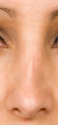
- The patient appears to have an “inverted V” deformity with collapse of the ULC’s
- Management would include revision septorhinoplasty with placement of bilateral spreader grafts
Review
- What are the most common causes of bony vault deformities?
- What are the most common Middle vault deformities?
- What are the most common Nasal tip deformities?
- How do you address bony vault deformities?
- How do you address Middle vault deformities?
- How do you address Nasal tip deformities?
- What are some of the Functional deformities that are seen after a rhinoplasty?
- How do you treat Nasal Functional deformities?
- Surowitz JB, Most S. Complications of Rhinoplasty. Facial Plast Surg Clin North Am. 2013 Nov;21(4):639-51
Learner must Sign In to access AAO-HNSF education activities.
- Annual Meeting Webcast (AMW):